
 Descargar número completo
 Download full issue
CITA ESTE TRABAJO
Sánchez Moreno S, Martínez Amate E. Hepatic angiosarcoma with elevated alpha-fetoprotein: a diagnostic challenge. RAPD 2025;48(3):112-113. DOI: 10.37352/2025483.4
Introduction
Elevated alpha-fetoprotein (AFP) is mainly associated with hepatocellular carcinoma (HCC), hepatoblastoma and germ cell tumors, but there may be other tumors that produce it, such as hepatic angiosarcoma, for whose diagnosis it is important to have a high index of suspicion.
Clinical Case
A 74-year-old patient with a history of metabolic syndrome and pneumoconiosis (due to occupational exposure to vinyl chloride) debuts with Budd-Chiari syndrome (BCS) with elevated alpha-fetoprotein (AFP: 6500 ng/ml). He presented a constitutional picture with dissociated cholestasis and radiological signs suggestive of chronic liver disease with thrombosis of the intrahepatic vena cava with extension to the suprahepatic vein.
Despite the high diagnostic suspicion of HCC, the dynamic liver study did not reveal compatible lesions and the liver biopsy was negative for malignancy. In addition, other tumors that can elevate AFP (teratoblastoma, germ cell tumor, Hodgkin's disease and gastric tumor) are ruled out. After initiation of anticoagulant treatment, the control angio-CT scan reveals a rapid radiological progression with extension of the described occupation to the right atrium in less than a month, with progressively increasing AFP levels (10,800 ng/ml). PET-CT (Figure 1) confirms moderate metabolic hyperuptake in the walls of the inferior vena cava from the right renal vein to the entrance into the right atrium, suggesting angiosarcoma as a first possibility. Combined surgical treatment (vascular and cardiac surgery) was proposed, but it was not possible because the patient died before the intervention.
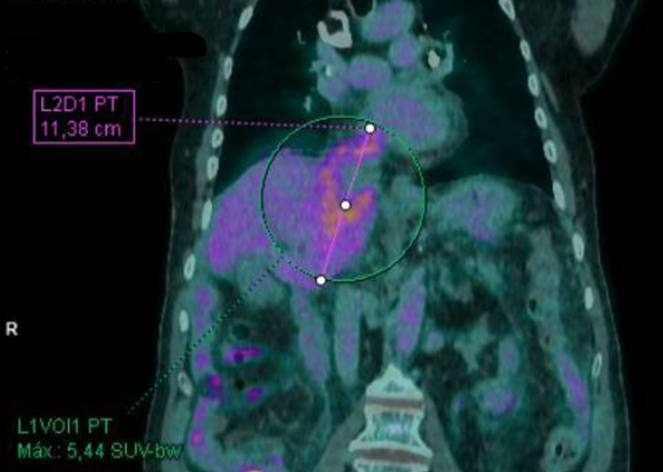
Figura 1
PET-CT. Moderate intensity metabolic hyperenhancement extending from the inferior vena cava and right renal vein to the right suprahepatic veins, reaching the entrance of the right atrium.
Despite not having a histological diagnosis (we do not have the necropsy consent), we believe it is a case of scientific interest due to its infrequency, unfavorable prognosis and the importance of establishing early suspicion.
Discussion
Vascular mesenchymal tumors are infrequent and have an insidious presentation with non-specific symptoms due to the formation of collateral circulation, and the diagnosis is made late in most cases[1]. Based on the evidence presented and the clinical course of the patient, we believe that the primary tumor may have been a hepatic angiosarcoma with massive secondary SBC, whose diagnosis was delayed given the initial suspicion of multicentric HCC based primarily on AFP levels. This represents the most common sarcoma in the liver and has been associated with exposure to carcinogens such as vinyl chloride and arsenic[2]. The other option proposed was leiomyosarcoma of the inferior vena cava of which there are some published cases[3]. However, the elevation of AFP is more indicative of the former, since its elevation is associated with HCC, hepatoblastoma and germ cell tumors, but it can be found characteristically elevated in other tumors such as hepatic angiosarcoma[4].
This case highlights the importance of excluding other rare vascular neoplasms in the presence of elevated AFP without clear evidence of HCC.