
 Descargar número completo
 Download full issue
CITE THIS WORK
Berdugo Hurtado F, Bailón Gaona MC, Moreno Barruecos M, Gutiérrez Holanda C. Acute pancreatitis and polycystic kidney disease, Is this relationship possible? RAPD 2025;48(1):34-35. DOI: 10.37352/2025481.5
Introduction
Autosomal dominant polycystic kidney disease (ADPKD) is a multisystemic disease with autosomal dominant inheritance with complete penetrance associated with mutation of the PKD1 and PKD2 genes. It is characterised by the presence of multiple bilateral renal cysts, as well as extrarenal manifestations that occur to varying degrees, with the development of cysts frequently at the hepatic level, known as hepatorenal polycystic disease, standing out at the digestive level. Like other digestive locations, cysts can be detected with low prevalence (9%) at the pancreatic level, which are almost always incidental findings[1],[2].
Clinical Case
A 66-year-old male with a history of chronic kidney disease associated with ADPKD who was admitted to our centre for a second episode of acute pancreatitis of an unidentified aetiology, ruling out in the anamnesis and with initial complementary tests (analysis and ultrasound) main causes of pancreatitis such as biliary lithiasis, toxic, hypercalcaemia, autoimmune, etc. At the analytical level, we highlight creatine of 1.8 mg/dl, dissociated cholestasis with gamma-glutamyl transferase of 650 U/L and alkaline phosphatase of 240 U/L, and amylase of 1450 U/L.
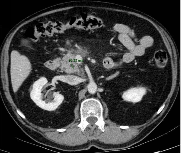
Given the patient's regular clinical evolution, abdominal computed tomography (CT) and magnetic resonance imaging (MRI) were performed, confirming the development of peripancreatic encapsulated necrosis at body level, highlighting the presence of several cystic formations in the head region and pancreatic uncinate process, the largest being 1 cm in size (Figure 1), causing minimal retrograde dilatation of the main pancreatic duct and the extrahepatic bile duct.
Figura 1
Axial section of abdominal CT scan with intravenous contrast. Cystic lesion 1cm in diameter at the level of the pancreatic uncinate process.
After conservative management of the process, the patient evolved favourably and was followed up on an outpatient basis with a control CT scan showing resolution of the necrosis, with persistence of a stable cyst at the level of the uncinate process without dilatation of the pancreatic duct or biliary tract.
Discussion
Incident cystic lesions at the pancreatic level are increasingly being diagnosed, with an estimated overall prevalence of between 13% and 18%, due to the increased use of more specific imaging tests such as CT or MRI3.[3].
In this case, and following the literature review carried out, we wish to show that pancreatic cystic lesions associated with ADPKD are rare, and the associated symptoms are even rarer. Isolated cases have been described of patients who developed episodes of chronic pancreatitis or cholangitis secondary to the obstructive process derived from these cysts[4],[5]. The first reported case of a patient with complicated chronic pancreatitis was published in 1998 by Malka et al[6].
In these cases, conservative therapy is the first option to be followed in patients with acute-chronic pancreatitis, reserving the surgical approach of targeted pancreatectomy depending on the location of the lesion causing the obstructive process for cases of recurrent pancreatitis with associated complications or episodes of recurrent cholangitis[4]-[6]. In our case, the patient showed a correct clinical evolution after applying conservative measures.